Contact Info
-
+54 9 11 4776.3814
¿What is Microtia?
The microtia is a congenital malformation (present from birth), and consists of a poor development of the ear, with many variants depending on the stage in which the formation of the ear stops during pregnancy.
It occurs in 1 in 6.000 or 10.000 live births depending on the area. In Asia it is more frequently, being in the order of 1 every 2.000 live births.
However, there are areas, especially in the highland sector, where the incidence increases, as in the north of Argentina, Peru, Bolivia and Ecuador, which has the highest Microtia rate in the world. It is not yet known why it happens, so parents do not have to blame themselves, they are not responsible for this. In 10% of patients, it occurs bilaterally.
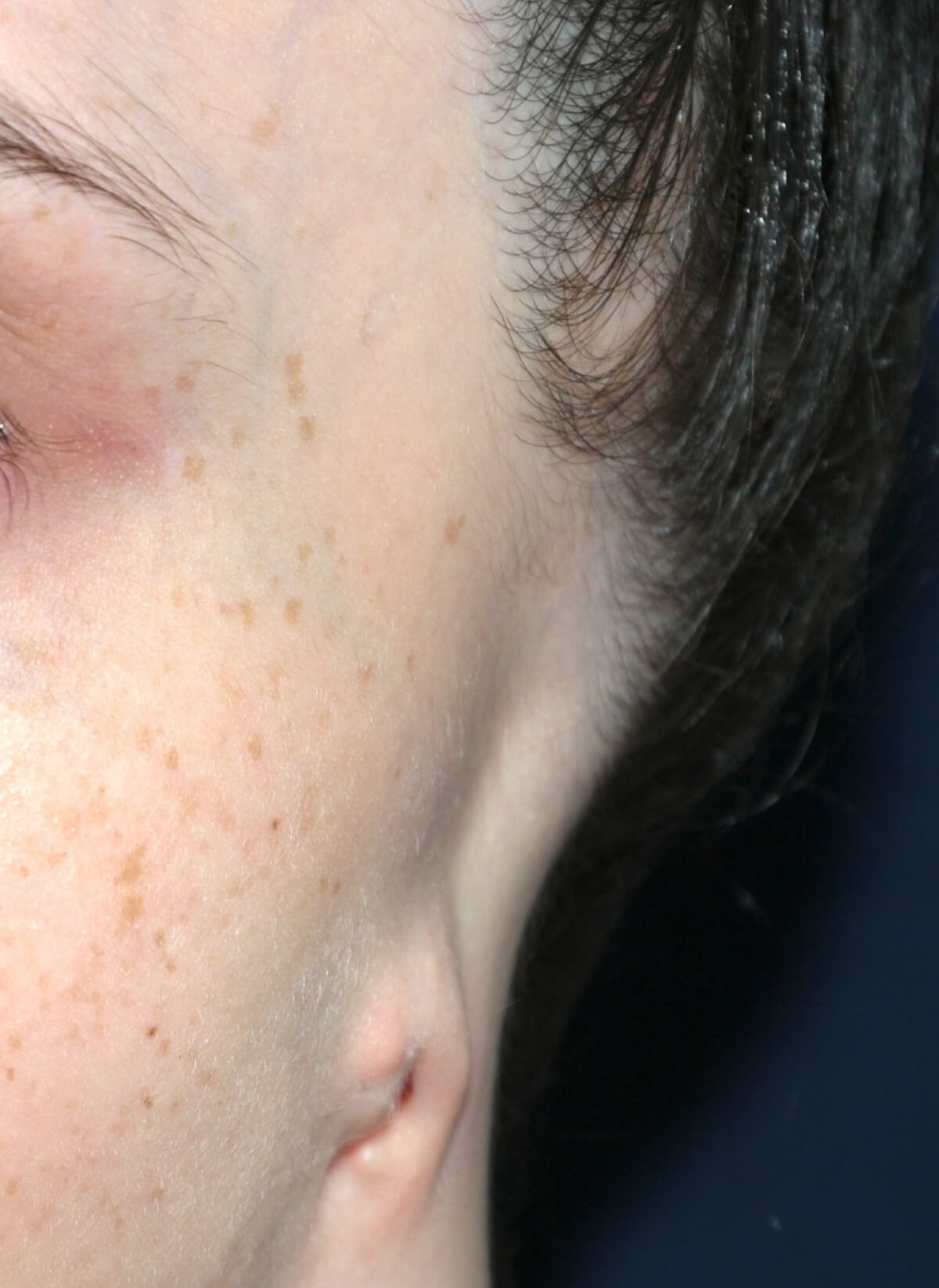
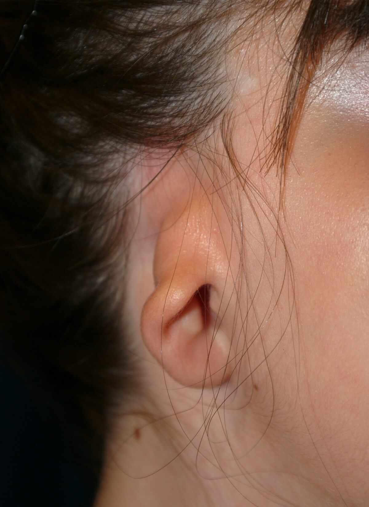
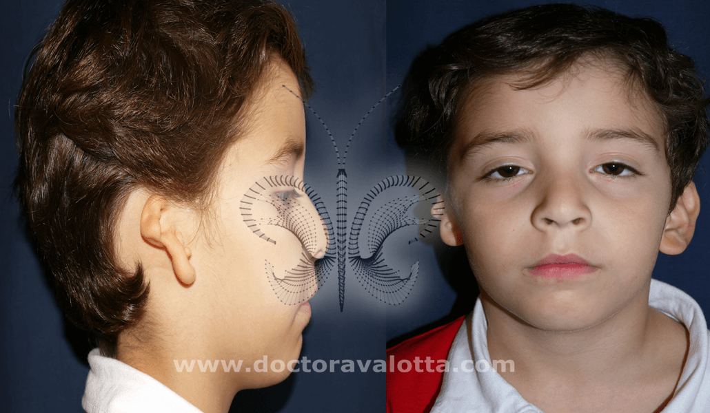
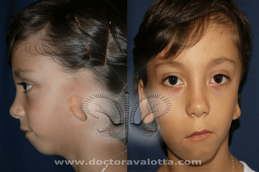
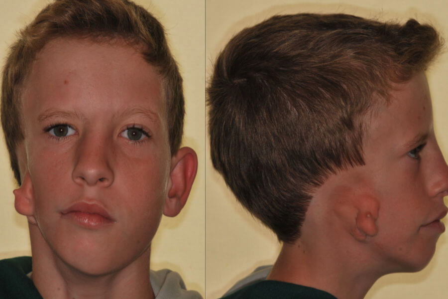
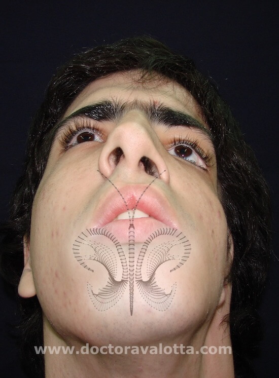
As the growth of the ear can stop at any stage, the malformations are very diverse, can go from a simple notch in the earlobe to the typical malformations we are accustomed to see. Within this spectrum there are many variants. A useful classification is that of Nagata, which divides Microtias into Lobular and Conchal types.
* Lobular type are the most common, the remnant is elongated and shapeless. In these cases we make a vertical incision to make the pocket and place the cartilage frame.
* In the Conchal type we usually use a horizontal incision. These cases often have tragus and antitragus.
* The complete absence of the ear is called ANOTIA.
Anotia
Microtia Conchal
Microtia Lobular
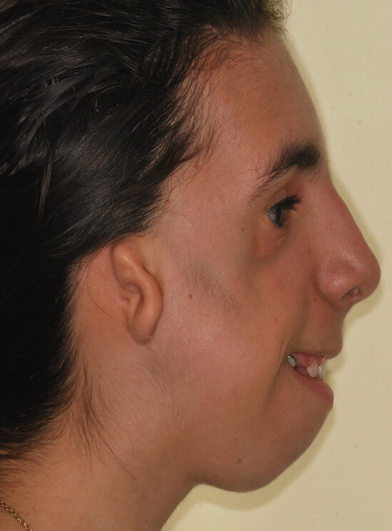
Hemicraneofacial Microsomics
n most cases Microtia occurs in isolation, without associated malformations. In other cases it is part of a more complex syndrome called Hemicraneofacial Microsomy. As the origin of the formation of the ear is common to other structures, in the microsomy the mandibular branch on the side of the microtia is shorter, which leads to a deviation of the bite. When it is very marked the lack of growth of the branch of the jaw can also affect the upper jaw and the orbit.
The remnant of the ear may also be displaced forward in more severe cases, and we may also find soft tissue atrophy, which is why fat grafts are very beneficial in these patients. When these children are born it is always important to discart kidney or heart problems, which are not very frequent but can occur. Hemicraneofacial microcopy may be mild, moderate, or severe.
ISOLATED MICROTIA
MILD MICROTIA

MODERATE MICROTIA
SEVERE MICROTIA
Goldenhar syndrome
It is the same as the microsomy, but renal, cardiac and cervical spine problems are more frequent. Unlike microsomy, this syndrome is genetic and is less frequent, occurring in 1 of every 25,000 live births. It has as typical characteristic an epibulbar cyst in the eye, which looks like a whitish lump. There may be bilateral forms.

Síndrome de Treacher Collins
It is a more complex, bilateral, genetic, autosomal dominant syndrome. This means that parents always transmit it to a greater or lesser degree, and there is no way to know what degree of deformity the child will have. Sometimes a parent has the syndrome in a very subtle way, and does not realize it. The syndrome is characterized by bilateral microtia, and a lack of bone and soft tissue in the cheek, with alterations in the eyelids. These malformations may be present or not, to a greater or lesser way. These children benefit greatly from fatty grafts performed at an early age.